NBEO Part 1 Anatomy and Physiology: Ocular and Neuro Topics
A focused guide to NBEO Part 1 anatomy and physiology: ocular anatomy, neuro-ophthalmic pathways, cranial nerve palsies, pupil defects, and visual pathway lesions.
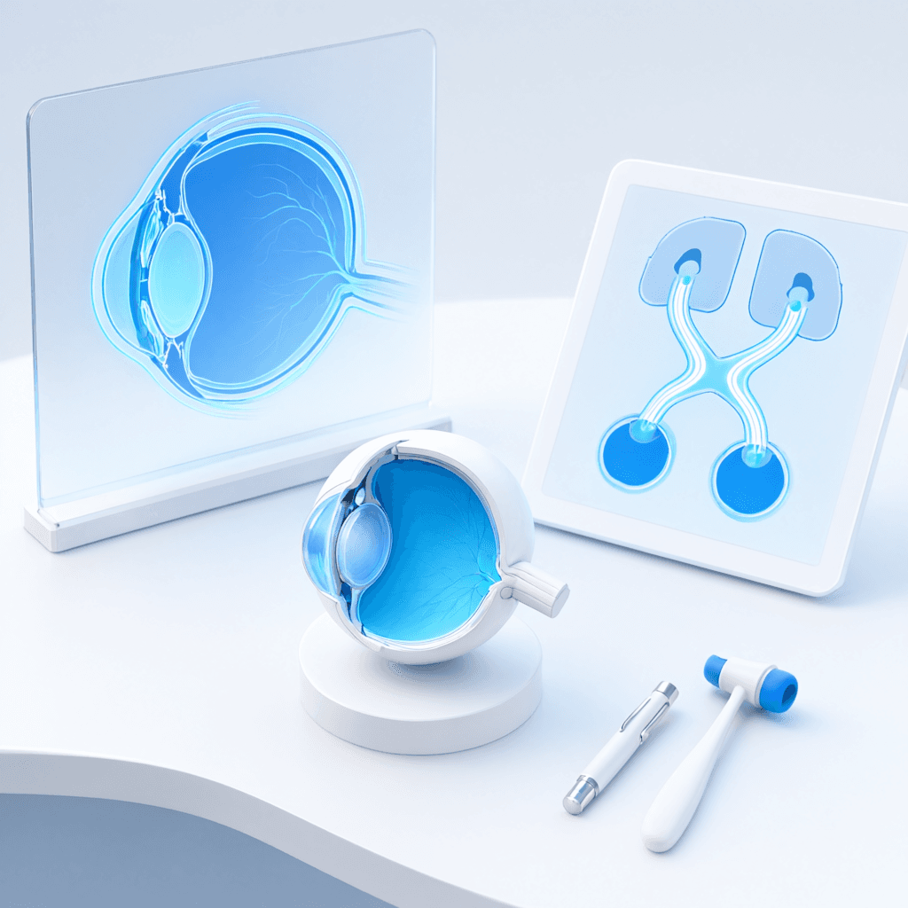
NBEO Part 1 Anatomy and Physiology: Ocular and Neuro Topics
Anatomy and physiology on NBEO Part 1 are tested in the context of clinical conditions, not as isolated facts. The most high-yield areas are ocular anatomy, neuro-ophthalmic pathways, cranial nerve workups, pupil defects, retinal layers, and the visual pathway. These topics connect directly to optics, pharmacology, and systemic disease, making them more valuable than their raw percentage weight suggests. Let's break down what actually matters and how to study it efficiently.
Anatomy is 12-18% of scored items on NBEO Part 1, and Biochemistry/Physiology is 10-14%. Those percentages are modest compared with Optics, but the overlap is huge. An item that asks you to localize a visual pathway lesion is testing anatomy, but it may also require you to interpret a visual field or choose a pharmacologic intervention. A cranial nerve palsy item tests anatomy, but the correct answer often depends on associated optics or systemic disease. This means anatomy and physiology should be studied as part of the integrated clinical picture, not as a separate memorization task.
Ocular Anatomy
A solid foundation in ocular anatomy is non-negotiable. You should be able to identify and describe the function of every major ocular structure. High-yield ocular anatomy topics include:
- Corneal layers and their clinical significance (epithelium, Bowman layer, stroma, Descemet membrane, endothelium).
- Iris and ciliary body anatomy relevant to accommodation and aqueous production.
- Lens anatomy, zonules, and cataract-related changes.
- Retinal layers and the blood-retinal barrier.
- Choroidal and scleral anatomy.
- Optic nerve head anatomy and cup-to-disc ratio considerations.
- Orbital bones, muscles, and vascular supply.
Retinal layer anatomy is especially important. Many students report items that require you to localize a lesion or drug effect to a specific retinal layer. Know the layers from photoreceptors to ganglion cells and how each layer contributes to visual processing.
Neuro-Ophthalmic Workups
Neuro-ophthalmic items are some of the most integrative on the exam. They require you to connect anatomy, physiology, and clinical presentation. High-yield topics include:
- Cranial nerve III, IV, and VI palsies: localization, associated findings, and expected eye positions.
- Internuclear ophthalmoplegia and its localizing value.
- Nystagmus types and their central versus peripheral causes.
- Optic neuritis, papilledema, and optic atrophy.
- Horner syndrome: localization, testing, and associated causes.
- Afferent pupillary defects and the swinging flashlight test.
A useful study technique is to draw the pupillary pathway and the visual pathway from retina to cortex. If you can trace the signal, you can localize the lesion.
Pupil Defects
Pupil items appear repeatedly. You must distinguish between:
- Relative afferent pupillary defect (RAPD): indicates asymmetric optic nerve or retinal disease.
- Effort pupillary defect: a normal finding during near effort.
- Anisocoria: physiologic versus pathologic, and the workup for each.
- Horner syndrome: ptosis, miosis, anhidrosis, and localization.
- Argyll Robertson pupil: light-near dissociation.
- Adie tonic pupil: light-near dissociation with segmental palsy.
For each pupil disorder, know the clinical findings, the anatomical localization, and the next step in evaluation. The swinging flashlight test is the key clinical maneuver for identifying an afferent defect.
The Visual Pathway
The visual pathway is another high-yield topic that connects anatomy to clinical localization. You should know:
- The path from photoreceptors through bipolar cells, ganglion cells, optic nerve, chiasm, optic tract, lateral geniculate nucleus, optic radiations, and visual cortex.
- The effects of lesions at each level (optic nerve, chiasm, tract, radiations, cortex).
- The corresponding visual field defects and any associated pupillary findings.
- The difference between contralateral and homonymous defects.
Practice localizing lesions based on visual field patterns and associated symptoms. This is a classic exam pattern that tests multiple levels of understanding at once.
Physiology Integration
Physiology items often connect to clinical function. High-yield physiology topics include:
- Aqueous humor dynamics and intraocular pressure regulation.
- Accommodation mechanism and the Helmholtz theory.
- Tear film physiology and dry eye pathophysiology.
- Corneal metabolism and the role of the endothelium.
- Lens metabolism and cataract formation.
- Phototransduction and dark adaptation.
These topics are most efficiently studied alongside their clinical counterparts. For example, study aqueous dynamics with glaucoma medications, and study tear film physiology with dry eye disease and pharmacology.
Common Clinical Scenarios on NBEO Part 1
The exam rarely asks you to name a structure in isolation. Instead, it presents a patient and expects you to connect anatomy to the presenting complaint. A few classic scenarios illustrate how ocular and neuroanatomy are tested together.
A patient with a painful, red eye and a mid-dilated pupil that does not react to light suggests acute angle-closure glaucoma. The item may ask you to identify the iris-corneal anatomy that explains the mechanism or the pharmacologic agents that alter aqueous dynamics. A patient with a sudden onset of vertical diplopia, especially when walking down stairs or reading, likely has a fourth cranial nerve palsy. The item may ask you to describe the superior oblique muscle, its innervation, or the head tilt test used to isolate the paretic muscle.
A patient with a relative afferent pupillary defect and a swollen optic nerve head raises the possibility of optic neuritis or anterior ischemic optic neuropathy. The correct answer may depend on whether you localize the problem to the optic nerve or the retinal ganglion cell layer. A patient with bitemporal hemianopsia has a chiasmal lesion, often from pituitary enlargement. The item may connect this visual field pattern to the anatomy of decussating nasal retinal fibers.
Practicing these scenarios helps you move from memorization to clinical reasoning. For each, identify the relevant anatomical structure, the expected clinical finding, and the related physiology or pharmacology. This integrated retrieval is exactly what NBEO Part 1 rewards.
Study Strategy
Anatomy and physiology are best learned through repeated active recall. Use diagrams, label blank images, and explain structures out loud. Flashcards work well for discrete facts like cranial nerve functions, retinal layers, and muscle innervations. For integrated topics like visual pathway lesions, draw the pathway and test yourself on the effects of lesions at each level.
If you want video crash courses, thousands of questions, and AI tools to drill your weakest anatomy topics, we'd love to help. Check out plans and pricing when you're ready.
Memory Techniques for Anatomy and Neuroanatomy
Anatomy requires memorizing a large number of structures, pathways, and relationships. These techniques can make the memorization more efficient and durable.
Draw it yourself. Passively looking at a diagram is weaker than drawing it from memory. Sketch the visual pathway, the pupillary pathway, and the orbit from memory, then check your drawing against a reference. The act of drawing builds spatial memory.
Use mnemonics for ordered lists. Cranial nerve functions, extraocular muscle innervations, and retinal layers are all easier to remember with mnemonics. Create your own if the standard ones do not stick, because personal mnemonics are more memorable.
Group by function rather than region. Instead of memorizing isolated structures, group them by what they do. For example, learn all the structures involved in aqueous production and outflow together, or all the muscles that elevate the eye together. Functional grouping creates logical connections.
Test yourself in both directions. For every pathway, practice naming the effect of a lesion and then naming the lesion that produces a given effect. The exam can ask either direction, so bidirectional retrieval is essential.
Use spaced repetition. Anatomical facts fade quickly without review. Add them to a spaced-repetition deck and review them daily, even when you are focused on other topics. Short, frequent reviews beat occasional long sessions.
Quick Review Checklist for Anatomy and Physiology
Use this checklist to make sure your anatomy and physiology preparation is clinically integrated.
- You can identify and describe the function of each major ocular structure.
- You can trace the visual pathway from retina to cortex and localize lesions.
- You can explain the pupillary pathway and distinguish afferent from efferent defects.
- You know the cranial nerves involved in eye movement and their clinical palsies.
- You understand aqueous humor dynamics, tear film physiology, and corneal metabolism.
- You can explain the visual cycle and lens metabolism in clinical terms.
- You have drawn the major pathways from memory at least once.
- You connect anatomy and physiology topics to their related optics, pharmacology, and systemic disease topics.
Anatomy and physiology are most valuable when studied as part of the clinical picture.
What This Means for You
Anatomy and physiology on NBEO Part 1 are not memorization contests. They are tools for understanding clinical presentations. When you study the visual pathway, you are also preparing for visual field and pharmacology questions. When you study cranial nerves, you are also preparing for optics and systemic health questions. Connect every anatomical structure to its clinical relevance, and you will retain more while answering more integrated exam items. Use diagrams, mnemonics, and spaced repetition, and always return to the clinical context.
Key Terms to Remember
- Visual pathway: The neural route from photoreceptors through the optic nerve, chiasm, tract, lateral geniculate nucleus, radiations, and visual cortex.
- Afferent pupillary defect: An asymmetry in the pupillary light response indicating disease of the retina or optic nerve on one side.
- Horner syndrome: A triad of ptosis, miosis, and anhidrosis caused by disruption of the sympathetic pathway to the eye and face.
- Cranial nerve palsy: A deficit in one of the cranial nerves controlling eye movement, commonly III, IV, or VI.
- Retinal layers: The layered structure of the retina from photoreceptors to retinal pigment epithelium, important for localizing lesions and drug effects.
- Aqueous humor dynamics: The production and outflow of aqueous humor that maintains intraocular pressure.
NBEO Part 1 anatomy and physiology are clinically integrated; focus on ocular anatomy, neuro-ophthalmic pathways, cranial nerve palsies, pupil defects, and visual pathway lesions. These topics reward understanding, not rote memorization — draw the pathways, talk through the scenarios, and connect every structure to its clinical picture. If you want a prep platform that emphasizes this integrated approach, we'd love to help you prepare. See plans and pricing when you're ready.
Ready to crack the NBEO Part 1?
Crack the Optometry Boards gives you 5 full-length NBEO Part 1 mock exams, 5,000+ exam-style questions, 30+ hours of video crash courses, 20,000+ flashcards, and a complete study engine: Adaptive Quiz Generator, Adaptive Flashcard Generator, AI Tutor, Homework Helper, YouTube Summarizer, and Study Schedule Creator. Everything is aligned to the official NBEO Part 1 blueprint and comes with unlimited tokens, unlimited credits, no throttling, unlimited access, and unlimited usage. No subscriptions, no usage caps.
No credit card required for the free trial. One-time payment for full access.
Related articles
NBEO Part 1 High-Yield Topics by Content Blueprint
Focus your NBEO Part 1 prep on the topics that appear most: optics, systemic health, neuro-ophthalmic anatomy, pharmacology, and the item-style traps that cost points.
Section GuidesNBEO Part 1 Optics Guide: Vergence, Magnification, and Binocular Vision
Master NBEO Part 1 optics with a focus on vergence, spectacle magnification, low vision, binocular vision, and contact lens calculations. Learn why conceptual understanding now beats formula memorization.
Section GuidesNBEO Part 1 Pharmacology and Systemic Health Review
Master NBEO Part 1 pharmacology and systemic health: drug classes, mechanisms, ocular side effects, and the systemic disease topics that make up the largest condition area on the blueprint.
Turn strategy into a passing NBEO Part 1 score
5 full-length NBEO Part 1 mock exams, 5,000+ questions, 30+ hours of video, and unlimited AI tools. Start today.
Unlock Everything